Nikon SMZ 18 Research Stereo Microscope
Request a Quote or DemoIntelligent yet affordable manual zoom stereomicroscope provides brilliant images and unparalleled uniformity in fluorescence intensity
Nikon’s SMZ18 research stereomicroscope combines macro and micro imaging in one manual instrument for convenient, affordable viewing and manipulation of single cells to whole organisms. A high-performance fly-eye lens provides crystal-clear fluorescent images, with uniform brightness across the entire field of view. Using enhanced epi-fluorescence technology, the SMZ18 is better able to detect excitation light than conventional fluorescent stereomicroscopes, for improved S/N ratios and bright, high-contrast images, even in the low magnification range. Integrated intelligence allows SMZ18 to save imaging parameters along with the captured image as a convenient data file.
Applications:
High-End Materials Science and Industrial Use
- Research and development activities across the full range of component parts and materials
- Production quality control functions for both routine and critical component applications
- Failure analysis studies to help determine, predict modes of failure, and verify applied solutions
- Component surface examination, crack detection, corrosion studies and coating efficiency
- Composite materials for aerospace, fabrics/textiles, building and construction goods
- Machine tool inspection for material integrity and features
- Optoelectronic devices by structures and component location
- Microelectronics, micro-electro mechanical systems (MEMS)
- Mobile phone components and technologies, as well as electrical shavers and fine watch manufacturing
- Medical devices including implants, prosthetics, and intra vascular systems
- Paint and coating technologies, inspecting both raw materials and in-situ coating integrity
- Art and antiquity conservation/restoration in both research and routine activities
Brightest Epi-Fluorescence Imaging
The SMZ18 combines fly-eye technology, which uniformly illuminates the entire field of view, and a high S/N ratio along with increased transmission for incredibly bright fluorescence, even at low magnifications. This also minimizes photobleaching and phototoxicity, thus reducing harm to live cells and organisms.
Flexible Architecture Allows Full Customization
A wide range of digital imaging capabilities means that users can customize the SMZ18 to best suit their research needs.
Improved Signal-to-Noise Ratios
Nikon’s newly developed optical system offers a drastic improvement in S/N ratio even at high magnifications.
The improved S/N ratio makes it possible to capture cell division, which is difficult using conventional stereomicroscopes, and samples with low excitation light.
Advanced Digital Imaging Capabilities
Access the Information you want Quickly and Easily
Easily obtain the information you need, such as Z drive position, zoom factor,
objective lens, filter cube, and LED DIA brightness by using the Digital Sight series and NIS-Elements or Digital Sight series DS-L3 together with the microscope. Sight series and NIS-Elements or Digital Sight series DS-L3 together with the microscope.
On-axis Imaging for Digital Images
Easily switch between stereo position (stereoscopic view) and mono position (on-axis view) when using the P2-RNI2 Intelligent Nosepiece by simply sliding the objective lens. Digital images with uncompromised clarity can be
captured using the mono position.
NIS-Elements Imaging Software
Nikon’s flagship, cross-platform imaging, software can now be used with Nikon’s newest stereomicroscope systems. NIS-Elements enables a wide range of advanced digital imaging capabilities, easily from a PC.
Digital Sight DS-L3 Digital Camera System
The DS-L3 is an easy-to-use high-definition, large touch-panel monitor that can be used to quickly capture images without a PC.
Access the Information you want Quickly and Easily
Easily obtain the information you need, such as Z drive position, zoom factor, objective lens, filter cube, and LED DIA brightness by using the Digital Sight series and NIS-Elements or Digital Sight series DS-L3 together with the microscope.
On-axis Imaging for Digital Images
Easily switch between stereo position (stereoscopic view) and mono position (on-axis view) when using the P2-RNI2 Intelligent Nosepiece by simply sliding the objective lens. Digital images with uncompromised clarity can be captured using the mono position.
NIS-Elements Imaging Software
Nikon’s flagship, cross-platform imaging, software can now be used with Nikon’s newest stereomicroscope systems. NIS-Elements enables a wide range of advanced digital imaging capabilities, easily from a PC.
Digital Sight DS-L3 Digital Camera System
The DS-L3 is an easy-to-use high-definition, large touch-panel monitor that can be used to quickly capture images without a PC.
Wide Range of Available Accessories
Nikon has improved ease of use by moving the controls to the front of the base, including the brightness adjustment dial and on/off switch.
- Fiber DIABase - Fiber DIA base features condenser lenses that can be switched between low and high magnifications. Furthermore, the Oblique Coherent Contrast (OCC) illumination system allows high contrast illumination.
- Slim Bases - The slimmer LED DIA Base and Plain Base help increase efficiency of sample manipulation by bringing the level of the sample closer to the table.
Focus Unit
The focus unit is combined with the base unit. Choose from either a manual or motorized focus unit.
SHR Plan Apo Series Objective Lenses
The SHR Plan Apo series features higher NA, wider field of view, and superior flatness and color aberration correction. These objective lenses can be seamlessly switched because all magnifications have the same parfocal distance. The new bayonet mount design allows lenses to be safely and easily removed.
Nosepiece / Focus Mount Adapter
There is one type of nosepiece that can mount two objective lenses and two types of focus mount adapters for use with a single objective lens.
Stage
The stage features an XY stroke of 6x4 inches (150mm x 100mm) and can be attached to any of the bases, making it effective for capturing large images when used in combination with the imaging software NIS-Elements. A sliding stage and tilting stage are also available.Controller.
Epi-Fluorescence Light Sets
- Motorized:The fluorescent turret can be operated using the remote control or imaging software NIS-Elements.
- Manual:An easy-to-use manual model for Nikon’s newly developed high performance epic-fluorescence attachment.
Fiber Illuminator Sets
- Flexible Double Arm Fiber Illumination Set:The direction and angle of illumination can be changed to suit the sample by making adjustments with these double arms. The fiber holder position can also be changed to obtain the optimal position for illuminating samples.
- Manual Epi-Fluorescence Light Set:This ring fiber illumination set features an episcopic illumination unit that effectively captures images (can be used with 1x and 0.5x objective lenses).
Coaxial Illuminator
The coaxial light illuminator makes it possible to view light reflected from the surface of a sample, which is ideal for shooting shadow-less images of thick samples.
Ring LED Illuminator
This ring fiber illumination set features an episcopic illumination unit that effectively captures images (can be used with 1x and 0.5x objective lenses).
Darkfield Observation Accessory
Darkfield viewing is possible simply by attaching the darkfield unit to the base.
Polarizing Observation Accessory
The analyzer is attached to the objective and the polarizer to the base or stand to enable polarized viewing.
| Zooming Body | |
| Optical System | Parallel-optics type (zooming type), apochromatic optical system |
| Zoom | Manual |
| Zoom Ratio | 18:1 |
| Zoom Range | 0.75–13.5x |
| Aperture Diaphragm | Zooming body built-in |
| Objectives NA, WD (mm) | |
| P2-SHR Plan Apo 2x | 0.3, 20 (with a correction ring for water 0 to 3mm in depth) |
| P2-SHR Plan Apo 1.6× | 0.24, 30 |
| P2-SHR Plan Apo 1× | 0.15, 60 |
| P2-SHR Plan Apo 0.5× | 0.075, 71 |
| Total Magnification (Using 10x eyepieces) | 3.75-270x (Depending on objective used) |
| Eyepieces (F.O.V. mm) | C-W 10xB(22) , C-W 15x(16) , C-W 20x(12.5) , C-W 30x(7) |
| Tubes (Eyepiece/ Port) | P2-TERG 100 Trinocular Tilting tube (100/0 : 0/100) P2-TERG 50 Trinocular Tilting tube (100/0 : 50/50) Inclination angle : 0°~30°
P2-TL100 Trinocular Tube L (100/0 : 0/100) Inclination angle : 15° |
| Focus Unit (Stroke from Objective's parfocal point) | P2-MFU Motorized Focus Unit (Up 96mm/Down 4mm) P2-FU Focus Unit (Up 97mm/Down 5mm) |
| Focus Mount Adapter/ Nosepiece | P2-FM Focus Mount Adapter P2-RNI2 Intelligent Nosepiece (2 objectives can be attached) |
| Bases/ Stand | P2-PB Plain Base P2-DBL LED Diascopic Illumination Base (OCC illuminator built-in) P2-DBF Fiber Diascopic Illumination Base P-PS32 Plain Stand P-DSF32 Fiber Diascopic Illumination Stand
P-DSL32 LED Diascopic Illumination Stand |
| Stages | P-SXY64 Stage , C-SSL Dia-sliding Stage , C-TRS Tilting Stage |
| Epi-Fluorescence Attachments | P2-EFLM Motorized Epi Fluorescence Attachment, P2-EFLI Epi Fluorescence Attachment |
| Epi-Fluorescence Light Sources | HG Precentered Fiber illuminator Intensilight C-HGFIE HG/C-HGFI HG (130W) |
| Episcopic Illuminators | P2-FIRL LED Ring Illumination Unit Use with Fiber light source: P2-CI Coaxial Epi Illuminator , P2-FIR Ring Fiber Illumination Unit
C-FDF Flexible Double Arm Fiber Illumination Unit |
| Episcopic Light Sources | C-FLED2 LED Light source for fiber illuminator |
| Observation Methods | (Episcopic) Coaxial Epi Illuminator, Epi Fluorescence Illuminator, Ring LED Illuminator (Diascopic) Simple Polarizing observation (with P2-POL Simple Polarizing Attachment), |
| Weight (approx.) | 30kg (Epi Fluorescence Attachment configuration with Trinocular Tilting Tube, Focus Unit, Intelligent Nosepiece, LED DIA base and Objectives 1x and 0.5x) |
| Power Consumption (approx.) | 10W (Epi Fluorescence Attachment configuration with Trinocular Tilting Tube, Focus Unit, Intelligent Nosepiece and LED DIA base) |
-

Nikon NIS-Elements Microscope Imaging Software
Provides intuitive tools for image capture, analysis, archiving, and image sharing.
More Information -

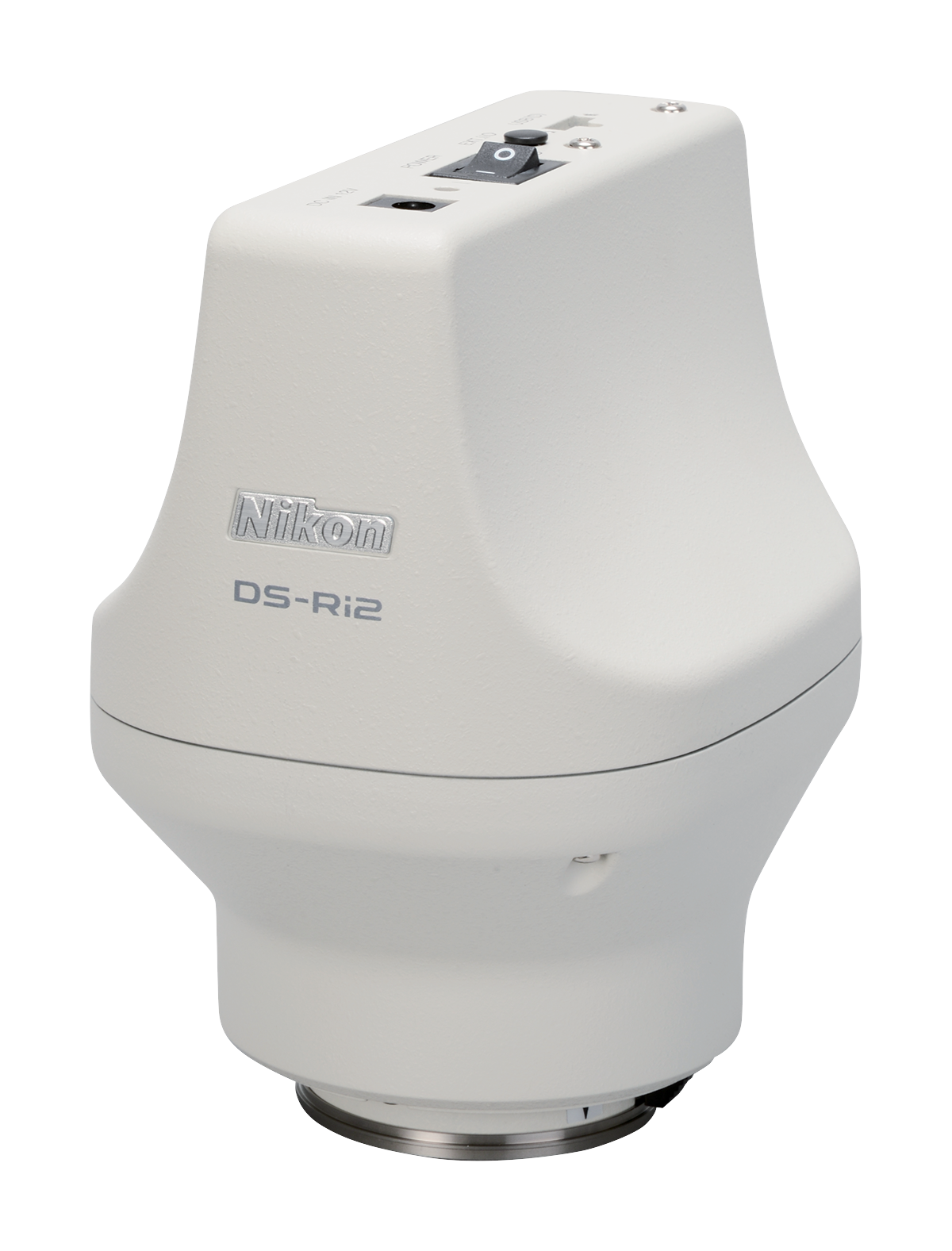
Discontinued: Nikon DS-Ri2 HD Digital Microscopy Camera
Replaced by: Digital Sight 10
More Information -

-

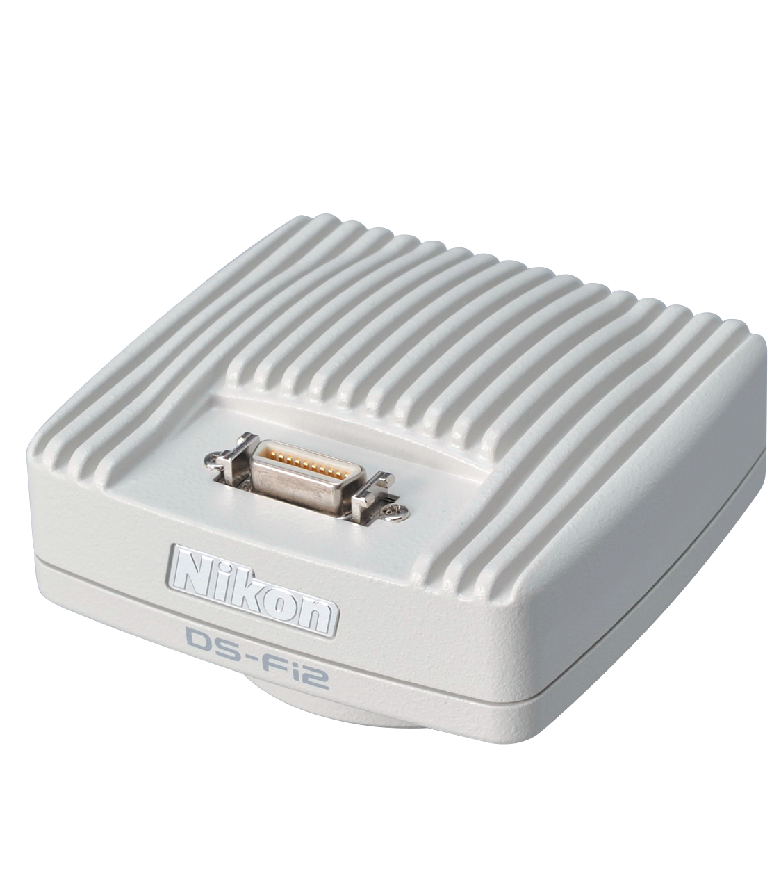
Discontinued: Nikon DS-Fi2 Microscope Camera
Replaced by: DS-Fi3 HD Color Microscope Camera
More Information -

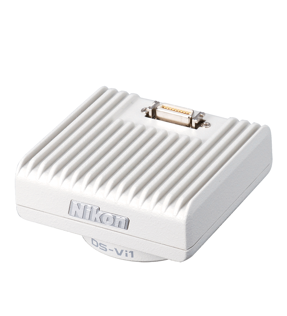
Discontinued: Nikon DS-Vi1 Color Microscope Camera
Replaced by: DS-Fi3 HD Color Microscope Camera
More Information


